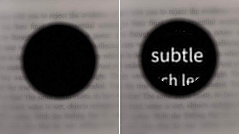
In a clinical trial of a wireless retinal prosthesis conducted by Stanford Medicine, people with advanced macular degeneration regained their vision enough to be able to read books and subway signs.
A tiny wireless chip implanted in the back of the eye and a pair of high-tech glasses have partially restored sight to people with an advanced form of age-related macular degeneration.In a clinical trial led by Stanford Medicine researchers and international collaborators, 27 of 32 participants regained their ability to read a year after receiving the device.
When digital features were provided by the device, such as zoom and contrast, some participants were able to read a very sharp vision equal to 20/42.
The results of the essay were published on October 20 at the New England Journal of Medicine.
The device, called PRIMA, developed at Stanford Medicine is the first prosthetic eye to restore functional vision to patients with severe visual impairment, giving them the ability to perceive shapes and patterns - also known as form vision.
"All previous attempts to provide vision with artificial devices focused primarily on light sensitivity, not actually the shape of vision," said Daniel Palankar, PhD, professor of ophthalmology and co-author of the paper."We provide vision first."
The other senior author is José-Alain Sachel, MD, professor of ophthalmology at the University of Pittsburgh School of Medicine.The lead author is Frank Holz, MD, professor of ophthalmology at the University of Bonn, Germany.
The two-part device consists of a small camera, mounted on a pair of glasses, that captures images and projects them in real time via infrared light onto a wireless chip in the eye.The chip converts images into electrical stimulation, effectively replacing natural photoreceptors that are damaged by the disease.
Prima is the culmination of years of development, research, animal testing and the first heart test.
Palankar first used such a device 20 years ago to treat eye diseases."We realized that the eye is transparent and needs to transmit information through light."He said.
"The device we envisioned in 2005 is now working well in patients."
To replace lost photoreceptors
The participants of the new trial gradually gradually gradually, gradually, gradually, more than 5 million people from the size of the world suffer from the condition, which is the cause of intolerance among minors.
Macular macular destroys the strong photoreceptors in the center of the retina, the thin layer and back into the electrical signal that is transmitted to the brain. But most patients retain some cells that allow visual vision, as well as neurons that contain information from photoreceptors.
The device we received is now working well in patients.
The new unit utilizes the preserved.
A 2-by-2 mm chip that receives images is implanted in the part of the retina where the photos are lost.The chip is analyzed from the glasses, unlike real photographers who only receive visible light, the chip is analyzed from the glasses.
"The projection was done in the infrared because we want to make sure that the rest of the photoreceptors are invisible outside the implant," said Palanker.
The design also means that patients with centric prosthetic vision can use natural vision in the center, which helps with orientation and navigation.
"It's important for them to be able to see the Prosthesis and the illusion at the same time, because they can integrate and use the vision fully," said Palanker.
Because the chip itself is programmable, which means it only needs light to generate power, it can use wireless and transfer via ReNena.The front sight is the satellite source and the cable that escapes the sight.
The new study included 38 patients over the age of 60 who had geographic atrophy due to age-related macular degeneration and vision worse than 20/320 in at least one eye.
Four to five weeks after the chip is implanted in one eye, the patient starts wearing glasses.Although some patients can make patterns right away, all patients' visual acuity improves over months of training.
"Achieving peak performance can take months of training, similar to what it takes for a cochlear implant to master a hearing aid," Palankar said.
Of the 32 patients who completed the one-year trial, 27 were able to read and 26 judged an improvement in visual acuity, defined as the ability to read at least two lines on the eye chart.On average, the participants' performance was better with 5 lines;one corrected in 12 lines.
Participants used the prosthesis in their daily lives to read books, food labels and subway signs.The glasses allowed them to adjust contrast and brightness and magnify up to 12 times.Two thirds reported moderate to high user satisfaction with the device.
Nineteen participants experienced eye effects, including eye contact), tears at the peripheral rate and solid collection.There is nothing wrong with them, and almost all of them are resolved within two months.
At the moment, the PRIMA device only offers black-and-white vision, with no shadows in between, but Palankar is soon developing software that enables the full range of grayscale.
"First on a patient's wish list is reading, and a close second is facial recognition," he said.– Grayscale is required for facial recognition.
And he's an engineer who delivers high-resolution thinking.Resolution is limited by the size of the pixels on the chip.Currently, pixels are 100 microns wide, with 378 pixels per chip.The new type, which has already been tested with mice, has pixels as small as a few microns, with 10,000 pixels per chip.
Palanker also wants to test the device for other types of blindness caused by missing photographs.
"This is the first version of the chip and has a relatively low resolution," he said."The next generation of chips will have smaller pixels and higher resolution. When paired with glasses, it will look more sophisticated."
Palanker said a chip with 20-micron pixels could give patients 20/80 vision. “But with electronic zoom they can get closer to 20/20.”
Palanker is a member of the WU TSAI Neurosciences Institute.
Researchers from the University of Bonn in Germany;Rothschild Hospital Foundation in France;Moorfield Eye Hospital and University College London;Ludwigshafen Academic Teaching Hospital;University of Rome II;Medical Center of the University of Lübeck Schleswig-Holstein;Red Cross Hospital and Claude Bernard University Lyon 1;San Giovanni Adolorata Hospital;Center Paradi Monticelli and University of Aix-Marseille;Crétil Community Hospital and Henri Mondor Hospital;Saarland National Hospital;University of Nantes;University Eye Hospital Tübingen;Münster University Medical Center;Bordeaux University Hospital;National Hospital 15-20;Erasmus University Medical Center;University of Ulm;Scientific Corporation;University of California, San Francisco;University of Washington;University of Pittsburgh School of Medicine;Sorbonne University contributed to this study.
This research was supported by a grant from the Science Corp.


